Archive for 2015
Wearable/Wireless | 3D Seismocardiography
 Researchers have been investigating the use of a promising, yet not entirely understood technique known as seismocardiography. This method takes advantage of natural vibrations produced by the cardiovascular system by recording with accelerometers, and using obtained data to make inferences about the state of health of the subject. Its use has shown promise as a noninvasive technique to measure heart health in both clinical and ambulatory environments. Researchers Paukkunen et al. have recently studied the three-dimensional vibration patterns of the cardiovascular system in an attempt to quantify them and make connections to the health of their subjects. To supplement their data, the researchers used a BIOPAC ECG amplifier and wireless respiration transducer to gain insight into the cardiovascular health of participants. Data was collected and analyzed from both a group of healthy subjects as well as a group of those affected by atrial flutter. The accelerometer and ECG/Respiration data was analyzed with AcqKnowledge, in an effort to understand more about the 3D vibration patterns and their use as indicators for disease. What the researchers found was that the data did differ significantly between the healthy subjects and those with heart flutter. How the data differed was in the relative location of these vibration events occurring in different parts of the cardiovascular system. By comparing to consistent cardiology data, the researchers were able to produce results that suggested that spatial distribution of seismocardiographic events. BIOPAC Systems offers these solutions and others for cardiology, with products designed for reliable, consistent data acquisition and analysis for wireless and wearable use in a variety of environments. This research sets the stage for further investigation into the potential use of seismocardiography to catch signs of heart disease easily and affordably, providing a new weapon for our long-lasting battle for cardiovascular health.
Surface EMG | Exercise and Muscle Fatigue
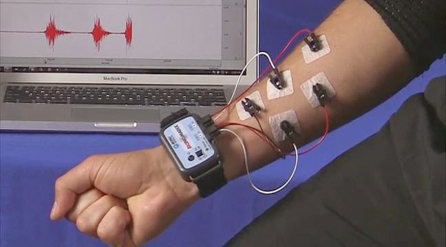
 Electromyography (EMG) has long been the clinical and research standard for skeletal muscle activity. Through manipulations of waveforms produced by electrical signals given off by muscle movement, researchers can gain insight into the mechanical properties of the muscular system. In exercise physiology, surface EMG is often used to study effort and usage dynamics of human muscles in an exercise setting. In a recent study by Jenkins et al, researchers observed the activity of the biceps and forearm during dumbbell curls between two groups. One group performed repetitions of curls with lighter weight (relative to their own 1- repetition maximum), and the other used heavier weight, both to the point of failure. The main difference here is that an exercise with a lighter weight can generally be repeated many more times than a heavier weight per set. There are differing views in the fitness community as to whether lifting heavier weights for fewer reps or lighter weights for many reps is more effective for optimal muscle activation. The researchers used a BIOPAC differential EMG amplifier and an MP150 system, combined with AcqKnowledge software to record data. The study shows, as a result of the surface EMG recordings, that there are significant differences in muscle activity in response to these differing methods of training. The total exercise volume, as a product of the weight lifted and reps performed, was similar between groups. However, a noticeable rise in EMG amplitude was recorded in those who performed more reps with light weights, suggesting greater muscle fatigue and activation in these individuals. These results conflict with previous studies performed with leg extensor resistance training, in which more muscle activation was observed in the heavier weight group. Thus the paper concludes that differences in muscle structure and blood flow may alter the effectiveness of different training methods across different muscle groups.
Wireless, Wearable | Quality of Life Technologies
There is a major concern growing in the medical community that the ratio of health workers to population size is decreasing. This means that the number of available doctors and medical professionals is starting to become too small to handle the number of people needing medical help. Technologies are therefore being created to help bridge the gap that is being created. These “Quality of Life Technologies” (QoLTs) have been developed to help monitor the health of people. While these technologies have been able to provide physiological support to individuals, the same could not be said for mental symptoms. If QoLTs could move into the realm of psychology and self-therapy, they could help improve the mood and quality of life for patients. A group of researchers from the Polytechnic University of Bucharest and the University of Lincoln recently published a paper that presents a machine learning approach for stress detection using wearable physiological amplifiers. To induce stress in participants, the researchers had them perform both a public speaking and cognitive task, which according to previous research these tasks caused the highest increase in measurable signals.
For their experimental setup, they used a BIOPAC BioNomadix BN-PPGED wireless transducer, hooked up to an MP150 data acquisition system, to record both EDA and PPG signals. They then used AcqKnowledge 4 software to extract both the PPG autocorrelation signal and Heart Rate Variability (HRV). Their results provided accurate stress detection in individuals. Their analysis marks a good starting point toward real-time mood detection, which could lead to people improving their quality of life. One way they could improve their experimental setup however, would be to use the BioNomadix Logger. This device allows for up to 24 hours of high quality data logging allowing the researchers to analyze a subject’s data from when they encountered stressful situations outside the lab.
For their experimental setup, they used a BIOPAC BioNomadix BN-PPGED wireless transducer, hooked up to an MP150 data acquisition system, to record both EDA and PPG signals. They then used AcqKnowledge 4 software to extract both the PPG autocorrelation signal and Heart Rate Variability (HRV). Their results provided accurate stress detection in individuals. Their analysis marks a good starting point toward real-time mood detection, which could lead to people improving their quality of life. One way they could improve their experimental setup however, would be to use the BioNomadix Logger. This device allows for up to 24 hours of high quality data logging allowing the researchers to analyze a subject’s data from when they encountered stressful situations outside the lab.
Data Logging | Understanding Social Fear Learning
Social
fear learning seems like a fairly straightforward subject. A person observes
another reacting or expressing through either verbal or nonverbal cues that a
stimulus makes them fearful or afraid. Surprisingly though, little is known
about how individuals modulate their perception of the threat. Researchers
hypothesized that understanding and shared emotional experiences with others
(empathy) play key roles in this, but there are a few investigations that
support it. Thus Andreas Olsson, Kibby McMahon, Goran Papenberg, Jamil Zak,
Niall Bolger, & Kevin N. Ochsner sought to study the role that empathy
plays in social fear learning. The experiment was set up across two stages;
one that tested manipulating empathy appraisals and the other individual
variability of trait empathy. Researchers enlisted a final sample of 47 men
and 53 women who attended Columbia University. The first stage had
participants receiving standard instructions that enhanced or decreased
empathy and underwent a fear learning procedure; the second had individuals
undergoing two observational learning procedures seeing whether the
participants expected to undergo the same learning as a demonstrator. During
the test stage, conditioned fear response was assessed through skin
conductance response (SCR) which was recorded from a BIOPAC MP150 system with
an EDA100C amplifier that monitors SCL and SCR data—BioNomadix wireless EDA or
Data Logger with EDA transmitter are viable
setup alternatives. SCR waveforms were analyzed with
AcqKnowledge software for off-line
analysis. The study found that subjects enhancing their empathy had the
strongest vicarious fear learning over the other groups. The results showed
that—especially in the strongly empathetic groups—a demonstrator’s expression
during the experiment tasks could serve as social unconditioned stimuli for
individuals to vicariously learn fear. Social fear learning thus depends on
both a person’s empathetic appraisal and their stable traits. Thus an
individual’s ability to learn fear from a social situation comes from not only
their inherent emotional state but also from their appraisal of how others
around them are reacting to the social
stimuli.
ECG Analysis | Physiological Benefits of Low-Altitude Tourism
 There
is a reason people call it “the Great Outdoors.” People often escape to nature
to relax and recharge from the stressors of suburban life. Wilderness tourism
has been and will probably continue to be very popular. Permit requests to
hike the Pacific Crest Trail increased so dramatically following Cheryl
Strayed’s book about hiking the trail and the film adaptation starring Reese
Witherspoon that the spike is known as “The ‘Wild’ effect.” While there are many enthusiasts
that spring for the harder challenge of higher altitude treks, many tourists
head for the lower altitude camp grounds and hiking trails. This allows
vacationers to experience the full benefits of wilderness tourism without the
knowledge required to battle various ailments of high altitude expeditions.
There is a certain comfort and relaxation that comes with a low altitude
nature getaway. People often credit this to the state of mind that comes with
being disconnected from modern life.
There
is a reason people call it “the Great Outdoors.” People often escape to nature
to relax and recharge from the stressors of suburban life. Wilderness tourism
has been and will probably continue to be very popular. Permit requests to
hike the Pacific Crest Trail increased so dramatically following Cheryl
Strayed’s book about hiking the trail and the film adaptation starring Reese
Witherspoon that the spike is known as “The ‘Wild’ effect.” While there are many enthusiasts
that spring for the harder challenge of higher altitude treks, many tourists
head for the lower altitude camp grounds and hiking trails. This allows
vacationers to experience the full benefits of wilderness tourism without the
knowledge required to battle various ailments of high altitude expeditions.
There is a certain comfort and relaxation that comes with a low altitude
nature getaway. People often credit this to the state of mind that comes with
being disconnected from modern life. A recent study sought to examine what physiological effects actually make people feel comfortable and relaxed at these low altitude camping areas. The authors Chen-Hsu Wang, Audrey Ming-Li Fan, Chen Lin and Cheng-Deng Kuo found that the real-effects of low-altitude tourism were not well documented. They decided to test three different low-altitude locations (30, 520 and 1080 MASL) and examined 49 healthy adults. Electrocardiographic signals were recorded using a BIOPAC MP System and analyzed using AcqKnowledge software. The study found that low-altitude wilderness tourism can lead to an increase in both Heart Rate (HR) and Blood Pressure (BP), and an increase in overall Heart Rate Variability (HRV). The paper notes that the greatest decrease in HR and BP and increase in HRV came around the 520 MASL mark. This shows that travel in low-altitude mountain areas may be good for physiological fitness in healthy adults for automatic nervous modulation and blood pressure regulation, especially in older individuals.
This experiment should prove to make that next vacation nature-oriented, whether it is to Yosemite, Big Sur or anywhere in between. This study shows that wilderness trips are not only good for the soul, but has positive physiological effects for your body as well.
Data Logging | Lumbar Multifidus (LM) Muscle
The lumbar multifidus (LM) muscle
is an important muscle that works to stabilize certain spinal segments as well
as control the extension moment of the lumbar spine. Studies have shown that
this muscle can be atrophied in people with chronic lower back pain. Physical
therapists thus frequently use lumbar extensor strengthening or stabilization
exercises for treatment of lower back pain. Researchers are still uncertain
about the influence of surface electromyographic (EMG) activity on lower back pain treatment outcomes. Recent
research has focused mostly on EMG levels during prone trunk extension (PTE) exercises and four-point
kneeling contralateral arm and leg lift (FPKAL) exercises. These recent
studies however have not focused on the selective activation of LM muscles
during lower back pain treatment exercises.
 Jun-Seok Kim, Min-Hyeok
Kang, Jun-Hyeok Jang, and Jae-Seop Oh thus sought to study
exactly that so as to provide an experimental study that established the
efficacy of the exercises as therapeutic treatment. The researchers gathered a
group of twenty healthy individuals without lower back pain who had not participated in lumbar strengthening or
stabilization exercises during
the previous six
months. Surface EMG data was
collected from the volunteers using a BIOPAC MP150 data acquisition and
analysis system as they performed the various exercises. The study found that selective
activation was higher during the FPKAL exercise than PTE, thus showing it is
the better and more effective way to treat lower back pain. While the
experiment provides good data for evaluating therapeutic exercises, future
evaluation in an actual physical therapy setting would prove
beneficial.
BIOPAC’s wireless BioNomadix Logger allows this type of
research to continue outside the laboratory. Subjects
who suffer lower back pain, for example, could wear the BIOPAC logging device when they are
performing PTE or FPKAL at home or during a therapeutic session.
The BioNomadix Logger’s portable
size and 24 hour data logging capability makes this type of surface EMG
recording outside the lab incredibly easy and would provide more insightful
evidence into effects of different therapeutic exercises.
Surface EMG | Musician’s Cramp
Focal hand dystonia, also known as “musician’s cramp,” is a movement disorder that causes involuntary flexing in the fingers, or finger cramps, when playing a musical instrument. This disorder poses a huge problem for professional musicians and in some cases can even threaten their careers. Many methods have been attempted to try and alleviate the ailment, but the most effective training method has been the “slow-down exercise” (SDE). This exercise, based on the fact that symptoms disappear when playing at a slow tempo, involves selecting a short passage that triggers the cramps then slowing the tempo down to where the musician can play without involuntary finger flexing. The same passage is then repeated over and over, gradually increasing the speed over time. While this method has helped improve symptoms, it was unclear what aspects of motor skills improved through SDE training.
Michiko Yoshie, Naotaka Sakai, Tatsuyuki Ohtsuki and Kazutoshi Kudo investigated how SDE affected motor performance, muscular activity, and somatosensation in a dystonic pianist. The study entitled “Slow-Down Exercise Reverses Sensorimotor Reorganization in Focal Hand Dystonia: A Case Study of a Pianist” tested a musician over a 12 month period as she underwent SDE training for 30 minutes a day, playing a specific passage that evoked the finger cramps most substantially. During the motor task, the musician’s surface EMG was recorded using EMG amplifiers and a BIOPAC MP Data Acquisition System. Throughout the rehabilitation process the musician improved her speed of key strokes and actually helped recover her normal motor and somatosensory functions. The researchers even found evidence that showed the brain had the capacity to reverse sensorimotor reorganization that was induced by the focal hand dystonia. The findings objectively show that SDE training not only improves effected people’s key strokes but helps to completely recover from the neurological disorder.
ECG Analysis | Body Dissatisfaction in University Attending Women
Weight
and body shape issues are a major concern amongst today’s general population,
especially young women. The pressure from outside forces to conform to a certain
body type, whether it is from advertisements or even their own social media
pages, is ever present. This causes a lot of women to harbor a high level of
body dissatisfaction which then internalizes aforementioned body shape
pressures. Mirror exposure has been used recently as a therapeutic technique to
reduce body dissatisfaction. Little is known, however, about what actually makes
this technique effective.
A recent study entitled “Body Dissatisfaction and
Mirror Exposure: Evidence for a Dissociation between Self-Report and
Physiological Responses in Highly Body-Dissatisfied Women” sought to study the
cognitive, mental and psychophysiological responses in women with different
levels of body dissatisfaction. Forty-two women attending University of Jaen were chosen to participate in the
study. The subjects were separated in to two groups, based on self-reported
criteria, into either the high-body dissatisfaction (HBD) or low-body
dissatisfaction (LBD) group. The participants were then asked to stand in front
of a mirror and directed to look at certain parts of their body (while wearing
beige underwear) while a BIOPAC MP150 system with an ECG amplifier continuously
recorded their physiological signals throughout the experiment. The researchers
then used AcqKnowledge software’s ECG analysis functions to obtain quantification of heart rate (HR) values. As
hypothesized, HBD women experienced more negative cognitive and mental emotions
than did LBD women. Conversely though, HBD women were found to have a reduced
physiological reaction (HR) than did LBD. The researchers hypothesized that this
might be due to HBD women’s development of a passive coping mechanism. Rather
than reacting with heightened senses to an upsetting or fearful situation, HBD
women react passively out of a possible sense of helplessness. Researchers also
felt that this could possibly be caused by HBD women performing more
self-inspections in the mirror than LBD women, but that the other explanation
was more probably. Although more research needs to be done, this study suggests
the possibility that eating behavior problems could stem from passive coping
mechanisms associated with body issues.
ECG Analysis | Physiological Changes in Response to Reporting
Physiological
responses can offer researchers key insights into the mental state of their
participants. Whereas human subjects can lie or misreport their emotions on a
self-report questionnaire, their physiological signals show the actual
truth. A quick rise in the recording
indicates a change in the subject’s emotions whether it be fear, anger, or
shame. Most studies concerning emotion shifts rely on heart rate data stemming
from ECG analysis and recording to see a participant’s reactions to the experimenters’ tests.
It is widely agreed that this is the true information that the analysis of the
ECG signal proves or disproves the study’s hypothesis. What if by simply
reporting on the emotion the researcher in fact influences changes in the
physiological response? That is what researchers Karim Kassam and Wendy Berry
Mendes sought to find out. They hypothesized that the awareness and conscious
assessment required by an individual for self-reporting of emotion may
significantly alter emotional processes. The researchers gathered one hundred
and twelve paid participants to take a series of tests designed to either induce
anger or shame (any individuals’ with depression or anxiety were excluded from
the study). Human subjects were either put into the anger, shame, or control
group and split by whether they were required to report their emotions during
the exam or not.
 Their physiological responses (heart rate, impedance
cardiography, cardiac output) were recorded using a BIOPAC MP150 Data Acquisition research system connected with
an ECG amplifier. The researchers found that in the anger group that
participant’s exhibited different physiological responses from those who were
not required to report. The shame condition however, seemed to show no
significant difference between the two groups (reporting and not reporting).
This study suggests that the act of reporting may have a substantial impact on
the body’s action to emotional situations. The data seems to point that
individuals who are provoked (anger) are likely to exhibit different
physiological responses when reporting. The knowledge that they will have to
explain their heightened emotions brings a rationale to an otherwise irrational
behavior. Shame on the other hand causes individuals to ruminate or
self-reflect, which would explain the little difference between the two
groups.
Wireless Physiology | Psychophysiological Measures of Emotion
Emotional reactions influence, and may help predict, our decisions and
offer valuable information for communication and neuromarketing researchers, but
emotion is difficult to measure explicitly. Emotional responses are complex
phenomena consisting of multiple components, including evaluation/appraisal,
subjective feeling, expression, and physiological reaction. This mix of
components is difficult to measure. Researchers can interview or survey
participants about their feelings—typical measures include traditional
Likert-type questions, open-ended questions, or pictorial scales—but
self-reporting doesn’t easily convey true or complete emotional response.
Self-reporting is further complicated by the fact that participations often
choose different terms to describe their feelings or respond that they feel
nothing. Blending self-assessment with physiological changes that reflect
visceral responses provides an unfiltered representation of
emotion.
 Significantly, EDA can provide time-stamped information for
moment-to-moment reaction measurement throughout a message/stimulus presentation
(such as an advertisement).Combining physiological data with self-reported data
helps provide a more complete, more accurate understanding of a participant’s
emotional reactions. Unobtrusive, wearable wireless physiology devices (such as BioNomadix
BN-PPGED from BIOPAC Systems, Inc.) can
provide continuous and precise measures of nervous system activity, such as EDA,
ECG, and RSP.
Sympathetic nervous system (SNS) activity provides objective data for
assessing emotional reactions. Electrodermal Activity (EDA) is a popular SNS
measure. EDA is basically an index of the electrical activity of the skin; sweat
glands in the skin are filled with tiny amounts of sweat and sweat contains ions
that conduct current, which can be detected and recorded. Increases in EDA
reflect increases in sympathetic nervous SNS activity. EDA is also referred to
as skin conductance (SCR, SCL, etc.) or galvanic skin response
(GSR).
 Significantly, EDA can provide time-stamped information for
moment-to-moment reaction measurement throughout a message/stimulus presentation
(such as an advertisement).Combining physiological data with self-reported data
helps provide a more complete, more accurate understanding of a participant’s
emotional reactions. Unobtrusive, wearable wireless physiology devices (such as BioNomadix
BN-PPGED from BIOPAC Systems, Inc.) can
provide continuous and precise measures of nervous system activity, such as EDA,
ECG, and RSP.
Read
a case study at “Hooked on a Feeling: Implicit Measurement of Emotion Improves Utility of Concept Testing.” Researchers conducted a message-testing study in which
they measured physiological
arousal (via EDA), emotional valence (via continuous rating dial data), and discrete
emotions (retrospectively reported emotional
reactions), among other measures. Researchers used a BIOPAC MP150 data
acquisition system and wireless EDA BioNomadix module to collect EDA while
participants viewed each ad, and a BIOPAC variable assessment transducer to
assess in-the-moment feelings of positivity or negativity. E-Prime was used to
allow for precise synchronization across stimuli presentation and data
collection.
Data Logging | Improvements in Wireless Wearables for Physiology Research
 Researchers have long recognized the need for a better tool for Electroencephalogram
(EEG) ambulatory monitoring. While many have come up with certain wired
solutions, there has not been a valid solution for long term logging or
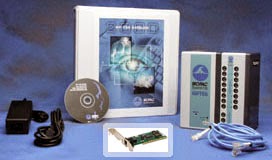
telemetry. BIOPAC Systems, Inc. has now introduced the brand new BioNomadix
Logger, a true wireless and wearable ambulatory monitoring device. The Logger
allows researchers the option for long term monitoring with its ability to
store data for later upload or it can also telemeter back to a computer for
live recording in a lab setting. The new BioNomadix logger truly allows
you to conduct physiology experiments anywhere and everywhere – you can log up
to 24 hours of high-quality data outside the lab and includes ECG,
EEG, EMG, EOG, EGG, EDA, Pulse, Respiration, Temperature, Cardiac Output, Heel
& Toe Strike, Clench Force, Accelerometer, & Goniometry signals. The
device is small in size, only about the size of your hand, making it easy for
subjects to take it with them in their everyday activities.
 The BioNomadix Logger fills the void where other more non-accessible devices could not. A study performed by researchers at the University of Minho aimed to create a wireless, wearable EEG ambulatory monitoring solution through a combination of other devices. They tested their tool against other devices (such as BIOPAC’s B-Alert X10) to measure the quality of the EEG signal. Although the researcher’s tool was able to record high quality data, it was bulky and could only be used on subjects in a lab. The BioNomadix Logger thus picks up where this study left off, allowing subjects to log data while performing everyday activities. The Logger’s small size also bypasses the bulky nature of other EEG ambulatory monitoring devices. The BioNomadix Logger thus represents a great step forward in long term wireless, wearable, ambulatory monitoring and data logging devices.
ECG Analysis: VLPs | Data Acquisition

An electrocardiogram (ECG or EKG) is a graphical recording of the changes occurring in the electrical potentials between different sites on the skin as a result of cardiac activity. The electrical activity of the heart is a sequence of depolarizations and repolarizations. Depolarization occurs when the cardiac cells, which are electrically polarized, lose their internal negativity. The spread of depolarization travels from cell to cell, producing a wave of depolarization across the entire heart. This wave represents a flow of electricity that can be detected by electrodes placed on the surface of the body. Once depolarization is complete, the cardiac cells are restored to their resting potential, a process called repolarization. This flow of energy takes on the form of the ECG wave, and is characterized by an initial P wave, followed by the QRS complex, and then the T wave. The P wave is associated with depolarization of the atria, the QRS complex is associated with depolarization of the ventricles, and the T wave with repolarization of the ventricles. Ventricular Late Potentials (VLPs, also called Ventricular Delayed Potentials) are small-amplitude, short-duration waves that occur after the QRS complex and are precursors to cardiac arrhythmias.
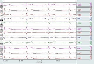
Use AcqKnowledge® software to apply signal averaging on the ECG signal to detect VLPs. To perform a VLP measurement on an ECG recording, use off-line averaging to trigger on the R-wave peaks and average the time delta of 209 ms before to 200 ms after the occurrence of each peak. AcqKnowledge measurement tools can calculate the duration and Root Mean Square (rms) values of the VLPs. AcqKnowledge also simplifies other ECG Analysis with powerful, fully automated routines for use post ECG recording: use the ECG averaging function to evaluate changes in the ECG complex before, during, and after exercise or dosing; perform heart rate variability (HRV) analysis; measure respiratory sinus arrhythmia (RSA); and more... http://www.biopac.com/ecg-cardiology
ECG Analysis of Putting Tournament
 An electrocardiogram (ECG; a.k.a.
EKG) recording can be extremely useful for analysis of a variety of
physiological studies. When combined with automated ECG analysis software,
researchers can identify ECG intervals, assess heart rate variability (HRV),
classify heart beats, and much more. ECG analysis results can be used with other
parameters to perform a complete physiological examination. Analyzing changes in ECG rhythms can provide
valuable insight into stress, arousal, and exercise research. A wide
range of physiological studies incorporate ECG results, such as a recent one
performed measuring cognitive workloads of participants during a competitive
golf putting tournament. The researchers set about to compare whether kinematic
or psychological factors were more important for participant’s putting
performance. Since putting requires more delicacy and precision on the part of
the golfer, they hypothesized that psychological factors would be just as
important --if not more-- than kinematic factors. The participants were divided
into three groups, arranged by skill level, and given the CSAI-2 test, an exam
that scored the participants’ self-confidence in the competitive environment.
The groups then performed in 8 tournaments of putting 2.1m from the hole. The
researchers used a BIOPAC ECG module to record and analyze heart rate and HRV,
which they ranked as either high or low. They found that the winners of the
tournament had a lower HRV frequency, which was associated with a lower mental
workload. There were also big differences in self-confidence scores on the
CSAI-2 between the winners and losers of the groups (specifically of the highest
skill level group). The results indicate that participants with lower mental
stress performed better, meaning that psychological factors are important to
putting ability. Although more research needs to be done, the results seem to
indicate that psychological factors seem to be more important to a golfer’s
short game.


